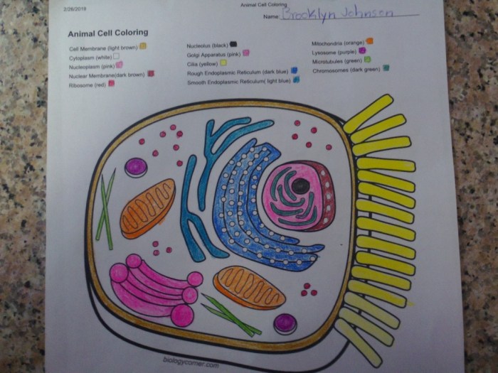
Introduction to Animal Cell Structure

Animal cell coloring key answers – Animal cells are the fundamental building blocks of animal tissues and organs. Understanding their structure is crucial to comprehending how animals function at a cellular level. These cells, unlike plant cells, lack a rigid cell wall and chloroplasts, resulting in a more flexible and adaptable structure. They are eukaryotic cells, meaning they possess a membrane-bound nucleus and other specialized organelles.Animal cells contain a variety of organelles, each with a specific function contributing to the overall cell’s life processes.
The efficient coordination of these organelles ensures the cell’s survival and proper functioning within a larger organism. Key differences exist between animal and plant cells, highlighting their unique adaptations to their respective environments.
Major Organelles and Their Functions
The following table Artikels the major organelles found in a typical animal cell and their respective roles. These organelles work together in a complex and coordinated manner to maintain cellular homeostasis and carry out essential life processes.
| Organelle | Description | Function | Analogy |
|---|---|---|---|
| Nucleus | The cell’s control center, containing the genetic material (DNA). It’s surrounded by a double membrane called the nuclear envelope. | Regulates gene expression, controls cell activities, and houses DNA. | The mayor’s office of a city, directing all operations. |
| Ribosomes | Small, granular structures, either free-floating in the cytoplasm or attached to the endoplasmic reticulum. | Synthesize proteins based on the genetic instructions from the nucleus. | Construction workers building proteins according to blueprints. |
| Endoplasmic Reticulum (ER) | A network of interconnected membranes extending throughout the cytoplasm. There are two types: rough ER (with ribosomes) and smooth ER (without ribosomes). | Rough ER synthesizes and modifies proteins; smooth ER synthesizes lipids and detoxifies substances. | A highway system transporting proteins and lipids throughout the cell. |
| Golgi Apparatus (Golgi Body) | A stack of flattened, membrane-bound sacs. | Processes, packages, and distributes proteins and lipids. | A post office sorting and shipping packages to their destinations. |
| Mitochondria | Double-membrane bound organelles often referred to as the “powerhouses” of the cell. | Generate ATP (energy) through cellular respiration. | Power plants generating energy for the city. |
| Lysosomes | Membrane-bound sacs containing digestive enzymes. | Break down waste materials and cellular debris. | Recycling and waste management center of the city. |
| Cytoplasm | The jelly-like substance filling the cell, surrounding the organelles. | Provides a medium for cellular reactions and supports organelles. | The city’s infrastructure, providing support and space for operations. |
| Cell Membrane | A selectively permeable membrane surrounding the cell. | Regulates the passage of substances into and out of the cell. | The city’s border control, regulating what enters and exits. |
Differences Between Plant and Animal Cells
Plant cells and animal cells share some common features as eukaryotic cells, but key differences exist. Plant cells possess a rigid cell wall providing structural support, chloroplasts for photosynthesis, and a large central vacuole for water storage and turgor pressure regulation. These features are absent in animal cells, reflecting their different ecological roles and physiological requirements. Animal cells, conversely, often have a more flexible shape and a greater range of motility.
For example, animal cells can move through chemotaxis or other means, unlike most plant cells.
Animal Cell Coloring Activities

Coloring activities are a valuable tool in teaching about animal cell structures. They engage students visually, helping them memorize the different organelles and their locations within the cell. This hands-on approach transforms a potentially abstract concept into a more concrete and memorable learning experience. The act of coloring reinforces the names and functions of each organelle, improving retention and understanding.
Common Organelles in Animal Cell Coloring Exercises
Five common organelles frequently featured in animal cell coloring exercises are the nucleus, cell membrane, cytoplasm, mitochondria, and ribosomes. Understanding their visual representation and typical coloring schemes is crucial for students completing these exercises.
- Nucleus: Typically colored dark purple or dark blue. This represents the control center of the cell, housing the genetic material (DNA).
- Cell Membrane: Often colored light blue or a light shade of green. It’s the outer boundary of the cell, regulating what enters and exits.
- Cytoplasm: Usually a light yellow or beige. This is the jelly-like substance filling the cell, where many cellular processes occur.
- Mitochondria: Commonly depicted in red or pink. These are the powerhouses of the cell, generating energy through cellular respiration.
- Ribosomes: Frequently colored dark green or brown. These are responsible for protein synthesis, crucial for the cell’s function.
Pedagogical Rationale for Color-Coding in Cell Structure Education
Color-coding in educational materials, particularly for complex biological structures like animal cells, serves a vital pedagogical purpose. The use of distinct colors for different organelles aids in visual differentiation and memorization. By associating specific colors with particular structures, students can quickly identify and recall the function and location of each organelle. This method enhances visual learning, making the abstract concept of cell structure more accessible and engaging for learners of all ages and learning styles.
For example, consistently associating the red color with mitochondria reinforces the understanding of their energy-producing role. Similarly, the dark purple nucleus readily becomes associated with its role as the cell’s control center. This visual association significantly improves recall and understanding during quizzes or examinations.
Interpreting Animal Cell Diagrams: Animal Cell Coloring Key Answers
Understanding animal cell diagrams is crucial for grasping the fundamentals of cell biology. Different representations of animal cells cater to varying learning styles and levels of biological knowledge. The ability to interpret these diagrams accurately is key to understanding cellular processes and functions.Different representations of animal cell diagrams exist, each with its own strengths and weaknesses for students at different learning stages.
Choosing the appropriate diagram type depends on the learner’s prior knowledge and the specific learning objectives.
Animal Cell Diagram Presentation Styles
Different presentation styles of animal cell diagrams cater to different learning levels and needs. Below are three examples illustrating this variety.
- Simplified Diagram: This type of diagram shows only the major organelles, such as the nucleus, cell membrane, cytoplasm, and mitochondria. It often uses simple shapes and labels to represent these components. For example, the nucleus might be represented as a large circle, and the mitochondria as smaller, oval shapes. The details of internal structures within these organelles are omitted.
- Detailed Diagram: A detailed diagram includes a more comprehensive representation of the cell’s organelles and their internal structures. It might show the rough and smooth endoplasmic reticulum, Golgi apparatus, ribosomes, lysosomes, and other organelles in more detail, possibly indicating their functions through labels or color-coding. This style might even include a scale to indicate the relative sizes of organelles.
- Microscopic Representation: This type of diagram attempts to visually mimic what an actual animal cell would look like under a powerful microscope. It might show the cell’s irregular shape and the relative positions of organelles as seen through a microscope. This often lacks distinct labeling of all components due to the complexity of the image, and interpretation relies heavily on prior knowledge.
So you’re hunting down those elusive animal cell coloring key answers? It’s amazing how much detail you can find in those tiny structures! Sometimes, though, I need a break from the microscopic world and switch to something a little more…visible, like checking out some awesome zoo animal coloring sheets for a fun change of pace. Then, refreshed and ready, I can tackle those animal cell diagrams again with renewed focus.
Advantages and Disadvantages of Different Presentation Styles
The effectiveness of each diagram style depends on the student’s background and the learning goal.
- Simplified Diagrams: Advantages: Easy to understand for beginners; quickly conveys the basic structure; suitable for introductory lessons. Disadvantages: Lacks detail; may oversimplify complex processes; not suitable for advanced learners.
- Detailed Diagrams: Advantages: Provides comprehensive information; suitable for advanced learners; facilitates understanding of complex cellular processes. Disadvantages: Can be overwhelming for beginners; requires prior knowledge of cell biology; may be difficult to interpret without adequate instruction.
- Microscopic Representations: Advantages: Provides a realistic visual representation; connects theory to real-world observations; useful for fostering critical thinking and interpretation skills. Disadvantages: Can be difficult to interpret without prior knowledge; may be too complex for beginners; requires understanding of microscopy techniques.
Advanced Animal Cell Components and their Representation
This section delves into less common organelles found within animal cells, exploring their functions and suggesting ways to visually represent them in a coloring exercise. Understanding these components provides a more complete picture of the cell’s intricate machinery.
While the nucleus, mitochondria, and ribosomes are frequently highlighted in cell diagrams, several other organelles play crucial roles in maintaining cellular function. Focusing on these less-common components enhances the educational value of a cell coloring activity by showcasing the cell’s complexity.
Three Less Common Animal Cell Organelles and their Functions
Three examples of less common organelles are centrosomes, lysosomes, and peroxisomes. Each plays a unique and vital role in the cell’s overall operation.
- Centrosomes: These are microtubule-organizing centers crucial for cell division. They organize the mitotic spindle, ensuring accurate chromosome segregation during cell replication. A malfunctioning centrosome can lead to errors in chromosome number, potentially causing diseases.
- Lysosomes: These are membrane-bound organelles containing hydrolytic enzymes. They function as the cell’s recycling centers, breaking down waste materials, cellular debris, and even invading pathogens. Lysosomal dysfunction can result in various storage diseases.
- Peroxisomes: These organelles are involved in various metabolic processes, primarily the breakdown of fatty acids and the detoxification of harmful substances. They also play a role in producing bile acids, essential for fat digestion. Peroxisome dysfunction can lead to a variety of metabolic disorders.
Visual Representation of Less Common Organelles in a Coloring Exercise
Visual representation of these organelles in a coloring exercise should be clear and distinguishable from more common organelles. Using distinct shapes, colors, and labels is essential for accurate depiction and comprehension.
- Centrosomes: Could be represented as a pair of small, darkly colored cylindrical structures near the nucleus, perhaps with radiating lines to indicate microtubules.
- Lysosomes: Could be depicted as small, oval-shaped organelles with a slightly darker interior color to suggest the presence of enzymes. They could be scattered throughout the cytoplasm.
- Peroxisomes: Could be shown as small, spherical organelles, with a distinct color and perhaps a slightly speckled interior to represent the enzymatic activity within.
Comparison of Common and Uncommon Organelles
The following table compares the function and visual representation of three common and three uncommon organelles, highlighting their differences for clarity in a coloring activity.
| Organelle | Function | Visual Representation |
|---|---|---|
| Nucleus | Houses genetic material; controls cell activities | Large, round structure; darkly colored; contains a visible nucleolus |
| Mitochondria | Produces ATP (energy); cellular respiration | Oval or rod-shaped structures; speckled or striped interior |
| Ribosomes | Protein synthesis | Small, dark dots scattered throughout the cytoplasm and on the rough endoplasmic reticulum |
| Centrosome | Microtubule organization; cell division | Pair of small, dark cylindrical structures near the nucleus; radiating lines |
| Lysosome | Waste breakdown; cellular recycling | Small, oval organelles; darker interior |
| Peroxisome | Fatty acid breakdown; detoxification | Small, spherical organelles; distinct color; possibly speckled interior |
Creating a Comprehensive Animal Cell Coloring Worksheet
This section details the creation of a comprehensive animal cell coloring worksheet, including a detailed diagram, an answer key, and suggestions for highlighting specific cellular processes using color. This activity is designed to enhance understanding of animal cell structure and function.Creating a detailed and engaging coloring worksheet requires careful planning and execution. The goal is to produce a visually appealing and informative tool that helps students learn the intricacies of the animal cell.
The worksheet should include a clear and accurate representation of the cell, with key organelles clearly labeled and easily identifiable. The answer key should be straightforward and easy to use.
Animal Cell Diagram Design
The animal cell diagram should be large enough to allow for easy coloring and labeling. It should accurately depict the relative sizes and positions of the various organelles. At a minimum, the diagram should include the following organelles: nucleus (containing the nucleolus), rough endoplasmic reticulum (RER), smooth endoplasmic reticulum (SER), Golgi apparatus, mitochondria, ribosomes, lysosomes, and the cell membrane.
Consider adding the centrosome for a more complete representation. Each organelle should be clearly labeled with a number or letter corresponding to the answer key. The style of the diagram can be simple and line-drawn or more detailed and realistic, depending on the target audience. For instance, the nucleus could be depicted as a large, central oval, the mitochondria as bean-shaped structures, and the endoplasmic reticulum as a network of interconnected membranes.
Ribosomes could be shown as small dots attached to the RER.
Answer Key Table
The answer key should be presented in a clear and concise table format. This ensures that students can easily check their work and identify any errors. The table should include a column for the organelle name and a column for the assigned color. For example:
| Organelle | Color |
|---|---|
| Nucleus | Light Purple |
| Nucleolus | Dark Purple |
| Rough Endoplasmic Reticulum (RER) | Light Blue |
| Smooth Endoplasmic Reticulum (SER) | Light Green |
| Golgi Apparatus | Yellow |
| Mitochondria | Red |
| Ribosomes | Dark Blue |
| Lysosomes | Orange |
| Cell Membrane | Brown |
Color-Coding Cellular Processes
Color can be used effectively to highlight specific cellular processes. For example, the pathway of protein synthesis can be emphasized by using a consistent color scheme. Ribosomes (dark blue) could be shown synthesizing proteins that then move through the RER (light blue) to the Golgi apparatus (yellow) for modification and packaging. The transport vesicles carrying these proteins could also be colored consistently (e.g., light orange).
This visual representation helps students understand the interconnectedness of organelles in carrying out essential cellular functions. Similarly, different shades of red could be used to represent different stages of mitochondrial respiration. This adds another layer of learning to the coloring activity.
Comparing Animal Cell Structures Across Species
Animal cells, while sharing a fundamental blueprint, exhibit fascinating variations across different species. These differences reflect the diverse adaptations required for survival in various environments and lifestyles. Comparing the cellular structures of different organisms reveals the remarkable adaptability of the basic animal cell design. This comparison focuses on human and insect cells, highlighting both commonalities and striking differences.
Human and insect cells, despite belonging to vastly different lineages, share the basic components of a eukaryotic animal cell. However, significant differences exist in the relative size, abundance, and even presence of certain organelles, reflecting their distinct physiological needs and evolutionary paths.
Organelle Size and Abundance Differences, Animal cell coloring key answers
The size and abundance of certain organelles vary significantly between human and insect cells. This reflects the different metabolic demands and functions of these cells.
- Mitochondria: Human cells generally possess a larger number of mitochondria compared to insect cells, reflecting higher energy demands of human tissues. This is especially noticeable in energy-intensive cells like muscle cells. Insect cells, while possessing mitochondria, often have fewer due to their generally lower metabolic rates and different energy strategies.
- Lysosomes: While both cell types contain lysosomes for waste degradation, the relative abundance and activity might differ based on dietary habits and cellular turnover rates. Human cells, with their complex dietary needs and longer lifespans, might have more active lysosomal activity compared to some insect cells.
- Endoplasmic Reticulum (ER): The extent of the ER network, both rough and smooth, can differ substantially. Human cells, with their diverse protein synthesis needs, often exhibit more extensive rough ER (studded with ribosomes) than some insect cells. Conversely, insect cells might show adaptations in smooth ER depending on their specific metabolic processes and detoxification needs.
Structural Variations in Cellular Components
Beyond the quantity of organelles, some structural differences also exist between human and insect cells.
- Cell Wall: A key distinction lies in the absence of a cell wall in animal cells. Human cells, like all animal cells, lack a rigid cell wall, allowing for flexibility and movement. Insect cells, however, may have extracellular matrices that provide structural support, though these are significantly different from the rigid cell walls of plant cells.
- Cell Junctions: The types and abundance of cell junctions, which mediate cell-to-cell communication and adhesion, vary considerably. Human cells utilize a range of junctions (tight junctions, gap junctions, desmosomes) for complex tissue organization. Insect cells may exhibit different junctional complexes tailored to their specific tissue architectures and physiological requirements.
- Vacuoles: While both cell types have vacuoles for storage, their size and function differ. Human cells generally possess smaller and more numerous vacuoles compared to some insect cells, which may have larger vacuoles for water storage or other specialized functions.
