Illustrating Beneficial and Harmful Bacteria

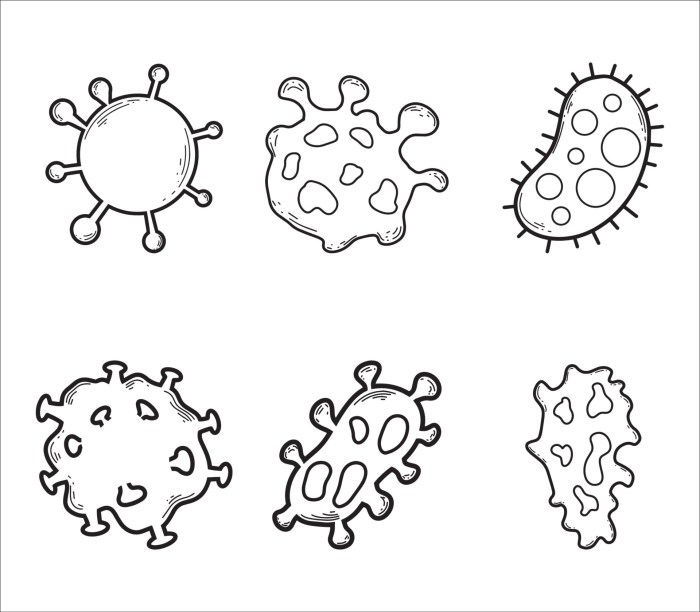
Bacteria burning drawing easy – Bacteria are single-celled microorganisms found everywhere on Earth. Some are beneficial to humans and the environment, while others are harmful and can cause disease. Understanding the differences between these types of bacteria is crucial for maintaining good health and understanding ecological processes. This section will illustrate these differences through visual representations and descriptions.
Beneficial Bacteria: Lactobacillus
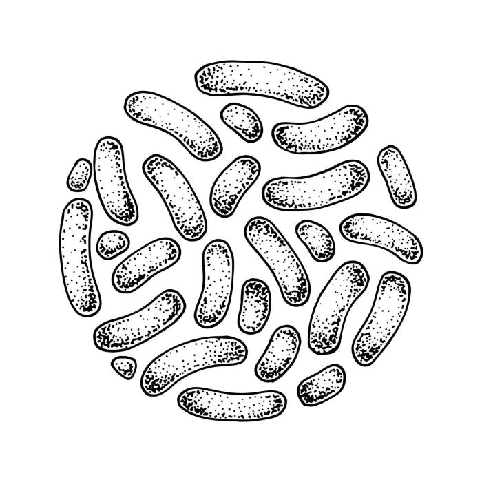
Lactobacillus is a genus of beneficial bacteria commonly found in yogurt, fermented foods, and the human gut. Imagine a drawing of a Lactobacillus bacterium. It would appear as a short, rod-shaped cell, slightly curved, with a smooth surface. Its shape is relatively simple. The cell might be depicted with a light pink or beige color.
There are no obvious external structures like flagella (for movement) visible. This is because Lactobacillus is generally non-motile. The text accompanying the drawing would explain that Lactobacillus helps in the digestion of food, producing lactic acid which inhibits the growth of harmful bacteria and improves gut health. It contributes to the overall balance of the gut microbiome, aiding in nutrient absorption and strengthening the immune system.
The drawing would clearly depict the rod-like shape, emphasizing its simplicity and lack of complex external features.
Harmful Bacteria: Escherichia coli (E. coli), Bacteria burning drawing easy
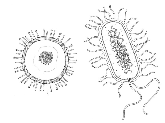
E. coli is a type of bacteria that typically resides in the intestines of humans and animals. While some strains are harmless, others can cause severe food poisoning and other illnesses. The drawing of E. coli would show a slightly longer, rod-shaped bacterium than the Lactobacillus, possibly with a more irregular shape or slight variations in thickness along its length.
Unlike the Lactobacillus, it might be depicted with a darker pink or reddish color, and potentially with small, hair-like structures called flagella extending from its surface, indicating its motility. These flagella would be clearly shown as thin, whip-like appendages enabling movement. The text accompanying the drawing would explain that pathogenic strains of E. coli produce toxins that can damage the intestinal lining, leading to diarrhea, vomiting, and abdominal cramps.
The drawing’s depiction of the flagella would highlight its ability to move freely within the body, facilitating its spread and colonization. The more irregular shape compared to the Lactobacillus could also suggest a more aggressive or adaptable nature.
Illustrating Bacterial Environments

Bacteria, the tiny prokaryotic organisms, exist in a vast array of environments, each presenting unique challenges and opportunities for survival and growth. Understanding these diverse habitats is crucial to appreciating the multifaceted roles bacteria play in the world around us. The following sections illustrate bacterial life in three distinct environments: soil, the human gut, and surfaces.
Bacteria in Soil
A drawing of bacteria in soil would depict a complex, three-dimensional environment. The soil itself would be represented as a mixture of mineral particles, organic matter (decomposing leaves, roots, etc.), and air pockets. Various types of bacteria would be shown interspersed throughout this matrix, some attached to soil particles, others free-living in the water films surrounding these particles.
Different shapes and sizes of bacteria (cocci, bacilli, spirilla) would be included to reflect the diversity of soil microbial communities. Some bacteria might be shown actively engaged in processes like decomposition, indicated by the presence of partially broken-down organic matter nearby. The overall image would convey the dense, interconnected nature of the soil bacterial ecosystem.
Bacteria in the Human Gut
A depiction of bacteria within the human gut would showcase the lining of the intestinal tract, with numerous bacterial cells adhering to the epithelial surface. Different bacterial species, represented by varying shapes and colors, would illustrate the diverse microbial community residing in the gut. Some bacteria might be shown interacting with each other, perhaps forming clusters or biofilms.
Other organisms, such as intestinal epithelial cells and immune cells, would also be included to highlight the complex interplay between the bacteria and their host. Beneficial bacteria might be depicted aiding in digestion or producing essential vitamins, while potentially harmful bacteria could be shown attempting to invade the intestinal lining. The overall impression would be one of a dynamic and densely populated ecosystem, crucial for human health.
Get ready to ignite your creativity with bacteria burning drawings! It’s surprisingly fun to depict these microscopic organisms engulfed in flames. For a change of pace, check out this awesome tutorial on animated drawing of two people easy – it’s a fantastic way to develop your animation skills! Then, return to your fiery bacteria masterpieces – you’ll be amazed at how much your drawing skills have improved!
Bacteria on a Surface: Biofilm Formation
A drawing illustrating bacteria on a surface forming a biofilm would show a layer of bacterial cells adhering to a solid substrate, such as a countertop or a tooth. The bacteria would be embedded within a self-produced extracellular polymeric substance (EPS) matrix, a sticky substance that holds the biofilm together and protects the bacteria from environmental stressors. The biofilm would likely exhibit a heterogeneous structure, with channels allowing for nutrient and waste exchange.
Individual bacterial cells would be shown at different stages of growth and development, highlighting the dynamic nature of biofilm formation. The drawing could also include depictions of other microorganisms, such as fungi or protozoa, that might be associated with the biofilm. The image would emphasize the complexity and resilience of bacterial biofilms.
Comparison of Bacterial Environments
The visual representations of bacteria in soil, the gut, and on surfaces highlight the diverse environments bacteria can inhabit and the adaptations they employ to thrive. Soil bacteria exist in a relatively open environment, interacting with a variety of organic and inorganic materials. Gut bacteria reside in a more enclosed and regulated environment, with a close relationship to the host organism.
Surface bacteria form complex biofilms, providing protection and facilitating interactions within the community. Despite these differences, all three environments showcase the fundamental characteristics of bacteria: their ability to adapt, their diverse morphologies, and their critical roles in various ecosystems.
Creating a Simple “Bacteria Burning” Illustration (Metaphorical)

This section will explain how to create simple drawings to metaphorically illustrate the effects of antibiotics and disinfectants on bacteria. We will use the visual metaphor of “burning” to represent the destruction of bacterial cells. This is a simplification, but it effectively conveys the concept to a basic level of understanding.The visual representation of “burning” is chosen to depict the rapid and destructive nature of these substances on bacteria.
The process is not literally burning in the sense of combustion, but the visual similarity helps to illustrate the effect of killing the bacteria. This approach uses readily understandable imagery to explain a complex biological process.
Antibiotic Action on Bacteria
To illustrate antibiotic action, draw several simple oval shapes representing bacteria. These ovals should be clustered together. Then, draw flames or a fiery effect engulfing and consuming some of the bacteria. The remaining bacteria should appear unaffected initially, but gradually more are consumed by the flames. This visually represents the process where antibiotics target and destroy specific bacteria.
The “flames” could be depicted as small, pointed shapes emanating from a central point, radiating outward to engulf the bacterial cells. The colour of the “flames” could be a vibrant orange or red, further emphasizing the destructive nature of the antibiotic. The surviving bacteria could be shown to be less active or diminished in size.
Disinfectant Action on Bacteria
For the disinfectant illustration, use a similar approach. Draw a cluster of bacterial ovals. This time, instead of flames, depict the disinfectant as a wave of intense, bright light, perhaps purple or blue, washing over the bacteria. The bacteria touched by this wave of light should immediately disintegrate or vanish, representing the rapid and broad-spectrum action of a disinfectant.
The light wave could be shown as a bright, solid wave front sweeping across the bacterial cluster, leaving no bacteria in its wake. This visual emphasizes the immediate effect of the disinfectant.
Answers to Common Questions: Bacteria Burning Drawing Easy
What materials do I need to draw bacteria?
You’ll primarily need paper, pencils (or pens), and colored pencils or markers (optional) for adding detail and color to your drawings.
Are there any specific software programs that can help with drawing bacteria?
While not necessary, drawing software like Adobe Illustrator or Procreate can assist with creating more detailed and polished illustrations. However, basic drawing tools are sufficient for this guide.
How can I make my bacterial drawings more realistic?
Research images of bacteria under a microscope. Observing real-life examples will help you accurately represent their shapes, sizes, and structures in your drawings.
Can I use this guide to draw specific types of bacteria?
Yes! The techniques and principles described here can be applied to drawing various bacteria. Research the specific bacteria you wish to illustrate for accurate representation.
